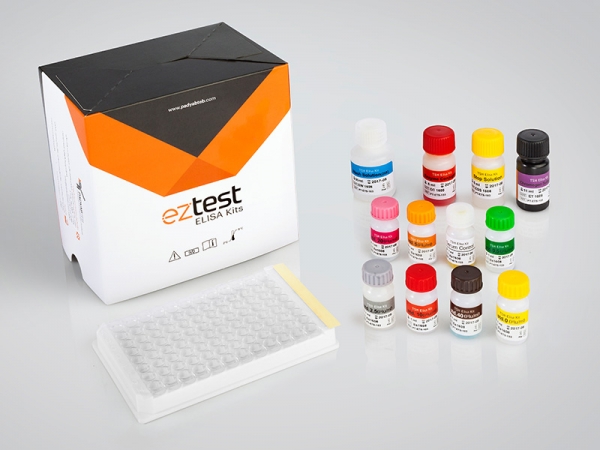
| Name | LH ELISA Test |
| Full name | Human LH ELISA Test Kit |
| Category Name | Fertility ELISA kits |
| Test | 96 |
| Method | ELISA method: Enzyme Linked Immunosorbent Assay |
| Principle | Sandwich Complex |
| Detection Range | 0-100 mIU/mL |
| Sample | 20µL Serum |
| Sensitivity | 1.0 mIU/mL |
| Total Time | ~60 min |
| Shelf Life | 12 Months from the manufacturing date |
LH ELISA kit description:
The Diagnostic Automation LH EIA test kit is for the quantitative determination of human luteinizing hormone (LH) concentration in human serum.
Materials provided with LH ELISA Test Kit:
1. Anti-LH Antibody-coated ELISA Microplate (96 wells)
2. enzyme conjugate reagent: 1 Vials contains anti-LH-HRP with preservative buffer, ready to use.(1 Vials, 12 ml)
3. standard set, contains 0, 5, 10, 25, 50, & 100 mlU/ml(standard 552/80, 2nd IS WHO) ready for use.(6 vials, 0.5 ml )
4. TMB Substrate: Hydrogen peroxide and tetra-methyl benzidine in preservative buffer, ready to use. (1 vial of 11ml)
5. Stop Solution: a normal hydrochloric acid (1 vial, 6 ml).
6. Concentrated washing buffer: PBS-Tween (1 vial 30ml) (20X)
7. Control serum: LH in serum with preservatives (1Vial, 0.5 ml)
Materials Required, not Provided:
1. Precision pipettes(20,50,100 µl)
2. Distilled or deionized water
3. EIA kit Microplate Washer
4. EIA kit Microplate Reader with a 450 nm, If possible, 630 nm as reference.
5. Absorbent paper
Introduction
LH (Luteinizing hormone) is a heterodimeric glycoprotein. Each monomeric unit is a glycoprotein molecule; one alpha and one beta subunit make the full, functional protein. The alpha subunits of LH, FSH, TSH, and hCG that are secreted from anterior pituitary are identical but The beta subunits vary and is responsible for the specificity of the interaction with the LH receptor. This hormone is produced by gonadotropic cells in the anterior pituitary gland. In both males and females, LH stimulates the secretion of sex steroids by the gonads. LH acts upon the Leydig cells of the testis in males. The Leydig cells produce testosterone. LH stimulates the secretion of estrogen in the ovaries. In women, ovulation of mature follicles on the ovary is induced by a large burst of LH secretion (preovulatory LH surge). Residual cells within ovulated follicles proliferate to form the corpora lutea that secrete the steroid hormones (progesterone and estradiol). In most mammals, progesterone is necessary for the maintenance of pregnancy and, LH is required for continued development and function of corpora lutea. In fact, the name of the LH is derived from this effect of luteinization from the ovarian follicles. The concentration determination of this hormone is important in fertility issue. This test is requested for assessing the pituitary function, including fertility issues, gonadal failure, issues during puberty, pituitary gland tumors, when the patient faces pregnancy difficulties and irregular or heavy menstrual periods, when it is thought that the patient has symptoms of pituitary or hypothalamus disorders, when there are symptoms of ovaries or testicles disease or when it is suspected that the child has delayed or early puberty as part of measures to infertility and pituitary or gonadal disorders.
Principle of the assay
LH test is designed based on immunoenzymometric assay. This assay is based on specific monoclonal antibody by the use of sandwich method. Anti- LH monoclonal antibody is used to conjugation with HRP enzyme for coating between the solid phase and an Anti- LH antibody. By addition of the sample serum, the LH molecule reacts with mentioned antibodies and sandwiches between bounded antibody on the solid phase and conjugated antibody. After an incubation period (45 minutes at room temperature), the sample is then decanted and the wells are washed to remove unbound-labeled antibody. An enzyme substrate-chromogen is added to the well and incubated for 15 minutes at room temperature, resulting in the development of a blue color. The addition of stop solution will stop the reaction and converts the color to yellow. The intensity is measured at 450 nm. The intensity of the yellow color is directly proportional to the concentration of LH in the sample.


